by Robert R. Rogers, DMD
How we can help treat snoring and apnea

The human pharynx is unique in the animal kingdom in that it is predisposed to collapse during sleep. In each and every one of us, the upper airway tends to diminish in size and shape to some degree from the relaxing influence of our nocturnal slumber. Most of us, fortunately, manage to breathe silently and effectively despite this ominous challenge to our well-being. This is not so for millions of others, however. Sleep-disordered breathing (snoring and obstructive sleep apnea) affects tens of millions of people worldwide. Obstructive sleep apnea (OSA) alone may afflict as many as 17% of Americans,2 a figure that is expected to approach 20% in the coming years due to the trend toward obesity.
On the most basic of levels, the human organism has but three critical needs for survival: eating, sleeping, and breathing. For those of us suffering from sleep-disordered breathing (SDB), two out of three of these requirements are in jeopardy every time our head hits the pillow. As sleeping and breathing are compromised night after night for years on end, the most recent research demonstrates that we are at greater risk for hypertension, heart attack, stroke, depression, and diabetes.3–8 In addition, it has been shown that patients with OSA are involved in traffic accidents two to three times more often than the general population9 and are at increased risk for injury in the workplace.10 Finally, the prevalence of undiagnosed sleep apnea in middle-aged adults suggests that untreated OSA may cause $3.4 billion in additional medical costs in the United States.11
The human pharynx is intimately involved with swallowing, breathing, and speaking; hence, it is called upon to be alternately stiff or flexible depending on the task at hand. The tendency, during sleep, for the upper airway to partially collapse (as in the case of snoring) or to completely obstruct (as with OSA) is multifactorial. Upper-airway patency is a delicate balancing act pitting pharyngeal anatomy and baseline muscle tone against the negative pressures created upon inhalation. During waking hours, patency is easily maintained. However, with the relaxing influence of sleep and the gravitational effects of the supine position, maintaining a clear airway becomes a titanic struggle for many people. Multiple stresses arise from intermittent airway occlusion, which include repetitive oxygen desaturation, carbon dioxide retention, and disrupted sleep from the arousals secondary to these blood gas discrepancies. As such, there are strong associations between OSA and the risk for cardiovascular disease and metabolic dysfunction.
Treatment modalities for managing sleep-disordered breathing include behavioral/lifestyle changes, positive airway pressure, surgery, and oral-appliance therapy. To date, none has proven universally effective with high patient acceptance, and therefore, health care providers are left with the task of mixing and matching patients to modalities.
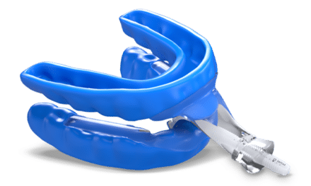
Over the past 15 years, properly trained dental professionals have emerged as key players in the treatment mix of this serious malady. By working closely with medical colleagues, dentists have been able to create and maintain upper airways in many patients to allow for adequate breathing and sleep. Therapy with oral appliances repositions and stabilizes the mandible, genioglossus, and hyoid bones. At the same time, the baseline muscle tone of much of the pharyngeal musculature is enhanced to diminish collapsibility during sleep.
In 1995, the American Sleep Disorders Association published a Review Paper and Practice Parameters. It focused on the scientific literature supporting the use of oral appliances in the treatment of snoring and OSA, and outlined the therapeutic protocols governing such use. This February, the same organization, renamed the American Academy of Sleep Medicine, published a revision of the Parameters in its journal, Sleep.12 In it, the use of oral appliances was found to be a necessary and effective mode of therapy to treat snoring and OSA. This scientific review focuses on several hundred studies and includes a preponderance of higher evidence levels than previously analyzed. And the practice parameters outline recommendations regarding the use of oral appliances as reflected in the outcomes of the studies reviewed.
It is wholly incumbent on any dental practitioner who treats snoring and OSA to be familiar with the entire contents of the Practice Parameters and to practice within the recommendations as published. Following are some pertinent highlights of the parameters:
“The presence or absence of OSA must be determined before initiating treatment with oral appliances (OAs) to identify those patients at risk due to complications of sleep apnea and to provide a baseline to establish the effectiveness of subsequent treatment. OAs are indicated for use in patients with mild to moderate OSA who prefer them to continuous positive airway pressure (CPAP) therapy, or who do not respond to, are not appropriate candidates for, or who fail treatment attempts with, CPAP. Until there is higher quality evidence to suggest efficacy, CPAP is indicated whenever possible for patients with severe OSA before considering OAs. Oral appliances should be fitted by qualified dental personnel who are trained and experienced in the overall care of oral health, the temporomandibular joint, dental occlusion, and associated oral structures. Dental management of patients with OAs should be overseen by practitioners who have taken serious training in sleep medicine and/or sleep-related breathing disorders with focused emphasis on the proper protocol for diagnosis, treatment, and follow-up. Follow-up polysomnography or an attended cardiorespiratory (Type III) sleep study is needed to verify efficacy, and may be needed when symptoms of OSA worsen or recur. Patients with OSA who are treated with oral appliances should return for follow-up office visits with the dental specialist at regular intervals to monitor patient adherence, to evaluate device deterioration or maladjustment, and to evaluate the health of the oral structures and the integrity of the occlusion. Regular follow-up is also needed to assess the patient for signs and symptoms of worsening OSA. Research to define patient characteristics more clearly for OA acceptance, success, and adherence is needed.”
Serious clinicians interested in comprehensive education and training in dental sleep medicine may contact the American Academy of Dental Sleep Medicine at www.dentalsleepmed.org. Board certification is offered by the American Board of Dental Sleep Medicine.
Prevention and Intervention
As the dental profession advances the application of oral appliances to manage the collapsible airway during sleep, we must not ignore the possibility of preventing the problem in the first place. As dental professionals, we are well-known for our commitment to prevention. In this regard, it is becoming apparent that the field of orthodontics may hold the key by virtue of its ability to manage facial form and airway integrity early on.
The AAO recommends orthodontic screening no later than 7 years of age. This recommendation derives from sound concepts and has served well in the past. However, more recent concern for upper-airway patency would seem to demand scrutiny much earlier. The age of 5 years has been suggested, but even by then the face has achieved most of its adult proportion. What, then, is an appropriate time to engage in the consideration of craniofacial morphology as it relates to sleep and breathing?
First, it may be prudent to consider the significance of this question and explore the implications of problematic sleeping and breathing in children. Habitual snoring affects 27% of all children,13 and even in the absence of frank, OSA has been associated with restless sleep and daytime sleepiness as well as neurobehavioral deficits such as behavioral hyperactivity and learning problems.14 As the partial upper-airway collapse (snoring) graduates to complete obstruction (OSA), comorbidity becomes more serious. The implications of OSA and the associated hypoxemia and sleep fragmentation in children are potentially complex. If left untreated or treated late, pediatric OSA may lead to significant morbidity affecting multiple target organs and systems, and such deleterious consequences may not be completely reversible despite appropriate treatment. The potential consequences of OSA in children include behavioral disturbances and learning deficits, pulmonary hypertension, systemic hypertension, and compromised somatic growth.15 In addition, pediatric OSA is associated with poor quality of life and increased health care utilization.16
It is abundantly clear that proper sleep and breathing are necessary for children to thrive. How and when to treat is of major importance. However, before addressing this dilemma, let us once again step back and consider some basic concepts such as the evolution of the modern human pharynx and the morphologic development of the upper airway.

As stated earlier, the human pharynx is unique in the animal kingdom because it is predisposed to collapse during sleep. Why is this so? To answer this question, one has to consider the function of the structures of the pharyngeal airway and compare the differences between other mammals and man. Postmortem dissections on many types of mammals reveal that the epiglottis extends up behind the soft palate to directly join the larynx to the nasopharynx (Figure 1, page 32). This provides a firm, uninterrupted air channel from the external nares, through the nasal cavities and nasopharynx, past the larynx, and down to the trachea and lungs. As such, no pharyngeal muscles were designed specifically to maintain upper-airway patency, since none were necessary. In addition, the tongue is located anteriorly, entirely within the oral cavity and separate from the pharynx, so it cannot impact the pharyngeal space at any time. This allows the animal to eat and breathe at the same time, preserving the sense of smell so necessary for survival.

It is interesting to note at this point that the pharyngeal anatomy of the human newborn and very young infants closely approximates the anatomy of the upper airway of non-human mammals.19 Specifically, the epiglottis and soft palate overlap and the tongue is completely anterior to the airway. No oropharynx, per se, is present. This allows for the simultaneous suckling of milk and breathing. By approximately 18 months of age, however, the laryngeal complex has migrated caudally, giving rise to the oropharynx, the ability to vocalize, and the concomitant predisposition to sleep-induced upper-airway collapse.
When to Screen
From very early on, the propensity for upper-airway collapse becomes a threat to breathing and, therefore, sleep. With this perspective, now is a good time to revisit the question of when to begin “orthodontic screening.” If we can include observation and monitoring of the upper airway as an integral part of the screening, then it would seem reasonable to begin within the first several years following birth. The ultimate goal would be to encourage craniofacial development that would maximize the potential for upper-airway patency during sleep throughout the life of the individual. This screening process would not necessarily be the exclusive domain of the orthodontist. A cooperative team approach that includes the parents, the pediatrician, the general dentist, and the ear, nose, and throat (ENT) surgeon would best serve the needs of the individual.
Although the newborn arrives with some genetic predisposition affecting upper-airway anatomy, it is well-documented that environmental and functional forces influence morphologic development. What can we do at the earliest that would benefit craniofacial development? Given the literature available, it seems reasonable to consider the benefits of breast-feeding over bottle-feeding, limit the use of commercial pacifiers, and control finger habits.
Breast-feeding has been shown to encourage proper development of the swallowing action of the tongue, good alignment of the teeth, and proper shaping of the hard palate.20, 21 Hultcrantz22 found that 6.2% of the children he studied snored every night by age 4, and another 18% snored when they were sick. While 60% of snorers used pacifiers, 35% of nonsnorers did. Breast-feeding has been the primary means of infant nourishment until relatively recent times. Palmer23 studied 600 prehistoric skulls from three different exhibition venues and noted a near absence of malocclusion. Of particular note were the broad hard palates and U-shaped, uncrowded arches in 98% of the specimens. Subsequent observation of more recent skulls from the bottle-feeding/pacifier era revealed a significant number of high, arched palates, narrow archforms, and crowded teeth. Indeed, Kushida et al24 have shown that a high palate and a narrow arch are good predictors of snoring and obstructive sleep apnea. Additionally, breast-feeding imparts certain immunological and antibiotic benefits that, when absent, may stimulate overgrowth of adenotonsillar tissue that can become a major obstructor in the young airway and can influence facial growth. Strong consideration should be given to adenotonsillectomy early on when frequent snoring and/or mouth-breathing is apparent.
As high, arched palates are seen with alarming frequency in our population, and are associated with mouth-breathing, ENT pathology, poor facial development, and sleep-disordered breathing, it seems reasonable for general dentists as well as medical practitioners to enlist the orthodontist as a key player in the creation of healthy faces that allow for healthy airways. Whether the arched palate is a result of genetic predisposition, maxillary compression during birth, improper molding from bottle-feeding or finger habits, or allergic adenotonsillar hypertrophy, the orthodontist can be very effective in orthopedically widening the maxilla. Many contemporary clinicians believe the ideal time for this may be between the ages of 4 and 8 years of age so as to prevent the related sequelae of the malformed hard palate. Often, however, we are forced to address problems later on in the older individual. In 1996, Cistulli25 showed that maxillary expansion was effective in resolving the sleep-disordered breathing events and daytime symptoms in an obese 22-year-old.
In Summary
Sleep-disordered breathing is a worldwide epidemic with health and economic consequences that range from annoying to deadly. Currently, treatment of snoring and obstructive sleep apnea calls upon a mix of modalities and a coordinated team of clinicians. Positive-airway pressure, surgery, and therapy with oral appliances are frequently effective in managing the sleep-induced unstable airway. However, common sense dictates that preventing disease in a natural way far outweighs any alternative that addresses the problem after it manifests. Those who place their health in our hands are best served if we can focus on the proper development of the upper airway immediately after birth and then work as a team to recognize and manage any abnormalities in breathing and facial morphology as early as possible.
Robert R. Rogers, DMD, is the director of dental medicine for St Barnabas Medical Center in Gibsonia, Pa. He is the founding president of the Academy of Dental Sleep Medicine (ADSM) and served again as president in 1995 and 1999. He is a diplomate of the American Board of Dental Sleep Medicine. Most recently, he was a member of the task force for the revision of the American Academy of Sleep Medicine Position Paper and Practice Parameters on Oral Appliance Therapy. He can be reached at [email protected].
References
1. Laitman J, et al. What the nose knows: New understandings of Neanderthal upper respiratory tract specializations. Proc Natl Acad Sci USA. 1996;93:10543–10545.
2. Young T, Peppard PE. Excess weight and sleep-disordered breathing. J Appl Physiol. 2005;1592–1599.
3. Krieger AC, Redeker NS. Obstructive sleep apnea syndrome: its relationship with hypertension. J Cardiovasc Nurs. 2002;17:1–11.
4. Smith R, Ronald J. What are obstructive sleep apnea patients being treated for prior to this diagnosis? Chest. 2002;121:164–172.
5. Moore T, Frannklin KA. Sleep-disordered breathing and coronary heart disease: long-term prognosis. Am J Respir Crit Care Med. 2001;164:1910–1913.
6. Shahar E, Whitney CW. Sleep-disordered breathing and cardiovascular disease: cross-sectional results of the Sleep Heart Health study. Am J Respir Crit Care Med. 2001;163:5–6, 19–25.
7. Saito T, Yoshikawa T. Sleep apnea in patients with acute myocardial infarction. Crit Care Med. 1991;19:938–941.
8. Reichmuth KJ, Austin D. Association of sleep apnea and type II diabetes: a population-based study. Am J Respir Crit Care Med. 2005;172:1590–1595.
9. Horstman S, Hess CW. Sleepiness related accidents in sleep apnea patients. Sleep. 2000;23:383–389.
10. Ulfberg J, Carter N. Sleep-disordered breathing and occupational accidents. Scand J Work Environ Health. 200;26:237–242.
11. Kapur V, Blough DK. The medical cost of undiagnosed OSA. Sleep. 1999;22:749–755.
12. Kushida CA, Morganthaler T, Littner M. Practice parameters for the treatment of snoring and obstructive sleep apnea with oral appliances: an update for 2005. Sleep. 2006;29(2): 240–243.
13. Arens R, Marcus CL. Pathophysiology of upper airway obstruction: a developmental perspective. Sleep. 2004;27(5):997–1019.
14. Fregosi RF, Quan SF. Ventilatory drive and the apnea-hypopnea index in six to twelve year old children. BMC Pulm Med. 2004;4:4.
15. Rosen CL, Larkin EK. Prevalence and risk factors for sleep-disordered breathing in 8 to 11 year old children. J Pediatr. 2003;142:383–389.
16. Ali NG, Pitson DJ. Snoring, sleep disturbance, and behavior in 4 to 5 year olds. Arch Dis Child. 1993;68:360–366.
17. Roberts D. Personal communication with Laurence Barsh. 1995.
18. Barsh LI. The origin of pharyngeal obstruction during sleep. Review article. Sleep and Breathing. 1999;3(1):17–21.
19. Laitman JT, Crelin ES. The function of the epiglottis in monkey and man. Yale J Biol Med. 1977;50:43–48.
20. Palmer B. The influence of Breastfeeding on the development of the oral cavity: a commentary. J Hum Lact. 1998;14(2):93–98.
21. Palmer B. The significance of the delivery system during infant feeding and nurturing. Australian Lactation Consultant Association News. 1996;7(1):26–29.
22. Hulcrantz E. The epidemiology of sleep-related breathing disorders in children. Int J Pediatr Otorhinolaryngol. 1995;32 (Supp):s63–s66.
23. Palmer B. The influence of breastfeeding on the development of the oral cavity: a commentary. J Hum Lact. 1998;14(2):93–98.
24. Kushida CA, Efron B. A predictive morphometric model for the obstructive sleep apnea syndrome. Ann Int Med. 1997;127:581–587.
25. Cistulli P. Treatment of snoring and obstructive sleep apnea by rapid maxillary expansion. Aust NZ I Med. 1996;26:428–429.


