New cone beams, cameras, and digital models
 |
Digital Models
3M Unitek, Monrovia, Calif, introduces its Lava™ Digital Models. The models are created using 3M’s direct impression Flash CT scanning process, which gives orthodontists the ability to make accurate diagnostic measurements with less variability than digital models made from plaster. The models can also be part of high-tech presentations during new-patient consultations. Lava Digital Models can be viewed in the free-of-charge Lava™ Treatment Management Portal (TMP) software that also provides an immediate ordering of iBraces™ Orthodontic Braces. The models are stored on 3M servers for 14 years.
3M Unitek
(888) 364-3577
www.3munitek.com
 |
Intraoral Camera
Gendex, Lake Zurich, Ill, introduces the GXC-300 intraoral camera. The GXC-300 provides visuals of the patient’s dental conditions that help dentists better explain treatment and, in turn, encourage patients to move forward with case acceptance. The portable GXC-300 uses true optics and a 4-LED lighting system to virtually eliminate image distortion, according to the company. The camera connects directly to a computer via high-speed USB cable—no docking station required. On and off activation of the camera is automatically controlled by placement in the holster. The GXC-300’s sliding, adjustable focus and quad-image capture buttons facilitate quick image capture.
Gendex
(888) 275-5286
www.gendex.com
 |
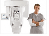
Cone Beam 3D System
PracticeWorks Systems LLC, Atlanta, introduces the KODAK 9500 cone beam 3D system for 3D imaging of dental and facial anatomy. The KODAK 9500 3D System enables dental professionals to produce 3D images ranging from dual jaw to full craniofacial images.
The system delivers anatomically correct 3D images down to 0.2 mm voxel size, and provides features such as face-to-face patient positioning to accommodate standing or seated patients—including wheelchair patients—of varying body types and sizes. Program selection with preprogrammed settings occurs directly on the system’s intuitive interface, simplifying patient setup and exam preparation.
The system also provides full-featured 3D visualization software, which includes an implant planning/simulation module and an implant library. Image output occurs in DICOM formats, supporting flexible output options including printing and customizable report templates.
The KODAK 9500 3D System captures dual jaw (9 cm x 15 cm) or all dento-maxillofacial anatomy (18.4 cm x 20.6 cm) in one acquisition for a wide range of clinical applications. With the additional ability to control patient dose through variable settings of mA and kV, dental professionals can manage the patient dose by focusing on their immediate clinical needs. This enables users to maximize image detail required for the specific application while managing patient dose.
PracticeWorks Systems LLC
(800) 944-6365
www.kodakdental.com