by Bradford Edgren, DDS, MS
3D imaging leads to an unusual discovery
 |
| Bradford Edgren, DDS, MS |
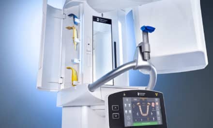
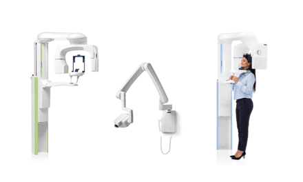
For an orthodontist, imaging provides an opportunity to delve into the patient’s present condition and prepare for proper growth and development. A 3D analysis provides the most detailed information for effective orthodontic treatment and diagnosis. Unfortunately, obtaining all of the angles that we need from 2D cephalometric (lateral and frontal head view) radiographs was often time-consuming and complicated. Since November 2007, when we integrated cone beam radiology into our practice, we got more than we anticipated—more precise diagnoses with some additional unusual discoveries.
 |
| A scan shows a mysterious itemin the patient’s ear. |
3D technology simplifies the process of obtaining lateral and frontal head views, plus a panoramic scan, all at once. With our 3D system, we don’t have to worry about magnification error or superimposition of other anatomical structures. Our i-CAT® has picked up undiagnosed supernumeraries, hidden and additional third molars, horizontally impacted canines, and horizontal root fractures—information that is vital to predictable treatment outcomes.
A discovery regarding one young patient made possible by our 3D imaging capability made us smile from ear to ear. A 6-1/2-year-old was referred by a general dentist for an orthodontic evaluation. She presented with severe crowding in her lower jaw and a tooth coming in completely sideways. Her medical history included tubes placed in her ears 4 years prior to her appointment with us. While evaluating her cone beam scan, I thought that my assistant had inadvertently forgotten to remove the child’s earrings, but she assured me that the child was not wearing earrings. However, the axial view on the MPR screen (which shows axial, lateral, and frontal views), showed a bright white foreign object visible halfway into the ear canal. I advised an immediate consultation with the ENT.
 |
| Mystery solved: the piece of pea gravel that had impeded the patient’s hearing. |
During the subsequent visit, the girl’s ear was inflamed and painful, but the ENT originally thought that the symptoms were a result of wax buildup. Our cone beam solved the mystery. Unable to remove the object during the office visit due to the child’s discomfort, the doctor determined the cause during surgery for already scheduled tonsil and adenoid removal. The ENT emerged from the operating room holding a plastic medicine bottle containing a piece of brown pea gravel, about 5 mm square by 2 mm thick, that could have gotten caught in her hair on the playground and subsequently pushed into her ear unnoticed by the preoccupied child. The ENT estimated that the rock must have been lodged in her canal for about a year. During that time, she was struggling in school, lip-reading because she was unable to hear out of her right ear. Since then, her mother ecstatically reports that her daughter is excelling in school.
|
Do you have a funny or heart- warming story from your practice that you would like to share? Contact Us with the details. |
So the strange but true headline to this story should read: “Patient’s hearing gets restored thanks to orthodontic exam.” My cone beam imaging helped this girl avoid potential long-term serious ramifications. Some unnoticed ear obstructions can result in severe, if not total hearing loss. Later that afternoon, as we looked at a 3D scan on another orthodontic patient, the image showed nothing unusual. Her mother said, “At least you don’t have rocks in your head!” If she only knew.
Bradford Edgren, DDS, MS, is in private practice in Greeley, Colo. He is a Diplomate of the ABO. He can be reached at