10/03/07
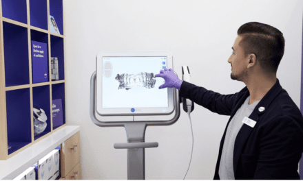
Imaging Sciences International’s Next Generation i-CAT® is now available for sale and installation. The i-CAT offers scan times at 5, 8.5, and 26 seconds. Its standard reconstruction takes less than 30 seconds, providing orthodontists instant data for patient diagnosis, treatment, and surgical predictability. Other features include a rotating, Amorphous Silicon Flat Panel Sensor for capturing small and extended fields of view.
“Imaging Sciences International set the bar for Cone Beam 3D imaging with the introduction of the i-CAT in 2004,” said Ed Marandola, president of Imaging Sciences. “Now, the Next Generation machine has raised the bar for dental imaging and treatment-planning. Forward-thinking dental practitioners have long relied on the i-CAT’s accurate, in-depth data, while patients appreciate the comfort and convenience of the in-office scans. The latest i-CAT continues our excellence in developing safe and effective treatment planning tools, and cements our leadership position within the industry.”
The i-CAT features typical file sizes of 50 megabytes so that orthodontists can share the images with referred specialists without worrying about lengthy downloads and special viewing software. It’s design features smooth, round corners for aesthetics and ergonomics. The sturdy and stable chair/head support mechanism reduces movement and optimizes image quality, while keeping patients comfortable. With the i-CAT, patients remain seated in an “open environment scan,” which captures the natural orientation of the anatomy. Once the data is captured, it’s transferred to a computer within minutes and displayed on an intuitive 3D mapping tool that allows orthodontists and technicians to easily format and select desired “slices” for immediate viewing. The footprint of the in-office i-CAT is 17-square feet.
For more information, contact Imaging Sciences.
[Business Wire, October 3, 2007]