by B. Giuliano Maino, MD, DDS
Miniscrews can achieve predictable results without patient cooperation
 |
Temporary skeletal anchorage in the form of miniscrews is becoming very popular and well-accepted by the orthodontic community because clinicians have experienced how useful miniscrews are in many clinical situations. In fact, by using miniscrews, we can move teeth in the desired direction with more predictability—sometimes within a shorter treatment period—and we can do it without patient compliance.
This can provide a huge advantage in comparison with the traditional treatment options, in which very often the patient’s cooperation is fundamental to obtain a good final result. A lack of cooperation during treatment can result in difficult situations with the loss of time and unsatisfactory end results often called compromised results. A lack of cooperation can also affect the end result right from the beginning. If a patient cannot provide adequate cooperation, the orthodontist is forced to choose a compromised treatment plan or a realistic treatment plan with acceptable results instead of an ideal treatment plan with optimum results.
Furthermore, there are several situations in which optimum results are difficult to achieve even in the presence of patient compliance: edentulous patients, periodontal patients, molar overeruption, control of vertical dimension, asymmetrical conditions, cant of the occlusal plane, and borderline cases.
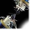
The use of skeletal anchorage today is facilitated by the fact that we can use miniscrews of small dimension (such as the Spider Screw® K1 by Ortho Technology Inc, Tampa, Fla) that can be placed in narrow spaces like the interproximal spaces.
The Spider Screw is easy to place because of its self-drilling and self-tapping design. Consequently, it can be inserted directly without a predrilled approach unless the cortical plate is thicker than 2 to 3 mm. This means that in the upper arch the miniscrew can usually be placed without predrilling, while in the mandible the cortical plate should be predrilled before insertion.
Case 1
This female patient had an unsatisfactory smile because of the excessive extrusion of the first and second molars and the second premolar on the upper left quadrant due to the loss of teeth in the opposite arch (Figures 1 and 1A). Molar overeruption can be a difficult task to correct with a conventional orthodontic approach because it requires the bonding of brackets in the entire arch and a long treatment time. The result can be unsatisfactory due to several factors, including extrusion of the adjacent teeth and limited intrusion of molars due to the poor efficiency of archwires in the final segment.
 |
 |
| Figure 1: Molars and premolar overeruption are due to the loss of teeth in the opposite arch. | Figure 1A: The patient smiling. |
Placing two miniscrews (1.5 mm in diameter, 10 mm in length in the tuberosity area, and 8 mm in length in the interproximal area) high in the vestibule permit the application of two nickel titanium coil springs of 100 to 150 g and obtain the desired amount of intrusion with high predictability (Figure 2A).
To prevent the buccal tipping of crowns when intruding upper molars, we can:
- Place a transpalatal bar and keep it apart from the palatal vault and from the palatal gingiva; or
- Use a miniscrew inserted on the palatal side (Figure 2B).
 |
 |
| Figure 2A: Miniscrews are placed in the vestibule to achieve intrusion. | Figure 2B: Miniscrew is placed on the palatal side. |
The use of a miniscrew on the palatal side permits the orthodontist to apply a force that can vary depending upon the size of the palatal root, and it will make the intrusive movement faster.1
Case 2
The control of vertical dimension is difficult in orthodontics. Many times, excessive vertical growth in the posterior part of the alveolar process without adequate growth of the condyle expresses itself in an open bite tendency.
Inserting one 1.5-mm miniscrew per side in the interproximal space between the first molar and the second premolar applies a vertical force to intrude the posterior teeth and decrease the posterior vertical dimension (Figures 3A, 3B, and 3C). A transpalatal bar was placed between the two first molars to prevent their buccal crown inclination (Figure 3D). The mandible underwent counterclockwise rotation, therefore closing the bite (Figures 4A and 4B, below).
 |
 |
| Figure 3A: A frontal view during molar intrusion with miniscrews. | Figure 3D: A transpalatal bar is placed to prevent buccal crown inclination of the molars. |
 |
 |
| Figures 3B and 3C: Miniscrews placed in the vestibule with elastic chain reduce vertical dimension. | |
 |
 |
| Figures 4A and 4B: Front view with bite closed and lateral view of the intruded molars. | |
Case 3
A Class II malocclusion needs patient cooperation to be treated successfully, especially in nonextraction cases. Patient compliance is usually greater at the beginning of treatment, while it decreases after the first 6 to 8 months.
Therefore, in the first phase of treatment, upper first molars have been distalized using a system that requires patient cooperation.
An acrylic cervical occipital (ACCO) anchorage appliance is combined with a cervical traction on the first molars. When the first molars reached an overcorrected Class I relationship, a miniscrew 1.5 mm in diameter and 10 mm in length was placed mesial to the upper first molars. A .016- x .022-inch stainless steel archwire with stops against the first molars and hooks crimped mesial to the canines was placed on the upper arch. According to the MGBM System,2 a .012-inch metal ligature was extended from each miniscrew to the hooks on the archwire to stabilize the overcorrected Class I position of the molars.
Canines and premolars were simultaneously retracted using hooks formed from stainless steel wire and inserted in the vertical slot of the brackets. Similarly crimpable hooks may be used and 150 g nickel titanium coil springs extended from the miniscrew to the premolars and canines (Figures 5A and B).
 |
 |
| Figures 5A and 5B: The miniscrews are in place. This second-phase lateral view shows canine and premolar retraction. | |
After the premolars attained a Class I position, the retraction of the incisors by means of the Bidimensional Technique3 sliding mechanics began. An .018- x .022-inch stainless steel wire with hooks distal to the lateral incisors and a piece of closed-coil spring between the second premolars and the first molars maintained the first molar in a stable position and prevented mesial movement of the molars so that there was no contact of the mesial roots and the miniscrews when they were inserted (Figures 6A, 6B, and 6C).
 |
 |
 |
| Figures 6A, 6B, and 6C: The third phase: incisor retraction. | ||
Stabilizing metal ligature wires were placed from the miniscrews to the canines, which were now in a Class I relationship. Traction in the form of nickel titanium coil springs of 300 g extended from the miniscrews to the hooks placed distal to the lateral incisors.
Total treatment time was 17 months.
B. Giuliano Maino, MD, DDS, is an active member of Angle Society of Europe. He is board certified by the IBO and EBO, and he serves as a visiting professor of orthodontics at Parma University. He is in private orthodontic practice in Vicenza, Italy, and he also does research in the area of skeletal anchorage using miniscrews. He can be reached at
References
- Maino BG, Bednar J, Pagin P, Mura P. The Spider Screw for skeletal anchorage. J Clin Orthod Dentofacial Orthop. 2003;2(37):90-97.
- Maino BG, Gianelly AA, Bednar J, Mura P, Maino G. MGBM System: New protocol for Class II nonextraction treatment without cooperation. Prog Orthod. 2007;8(1):130-143.
- Gianelly AA. Bidimensional Technique. Theory and Practice. New York: GAC International Inc; 2000.